- Tel: 858.663.9055
 Email: info@nsjbio.com
- Tel: 858.663.9055
- Email: info@nsjbio.com
Autophagy Antibodies are essential tools for studying the highly conserved cellular process of autophagy, which maintains homeostasis by degrading and recycling cytoplasmic material. Through autophagy, cells remove damaged organelles, misfolded proteins, and intracellular pathogens, ensuring survival during stress and nutrient deprivation.
Key autophagy markers include LC3 (microtubule-associated protein 1A/1B-light chain 3), Beclin-1, p62/SQSTM1, ATG proteins, and LAMP family proteins. By targeting these proteins, Autophagy Antibodies allow researchers to monitor autophagosome formation, flux, and lysosomal degradation. Because autophagy intersects with cancer, neurodegeneration, infection, and aging, the Autophagy Antibodies portfolio has broad applications in basic science and translational research.
NSJ Bioreagents provides Autophagy Antibodies validated for immunohistochemistry, western blotting, immunofluorescence, flow cytometry, and ELISA. Each Autophagy Antibody undergoes rigorous quality control to ensure specificity, reproducibility, and performance across multiple assay systems.
By choosing Autophagy Antibodies from NSJ Bioreagents, scientists gain reagents optimized for reliability and clarity. Our antibodies provide strong detection of autophagy proteins in tissues and cell lines, reproducible quantification of autophagy flux, and dependable performance across discovery and diagnostic platforms. Comprehensive datasheets, validated controls, and optimized protocols ensure reproducibility in laboratory and translational workflows.
The Autophagy Antibodies collection supports diverse applications in cell biology, pathology, and translational medicine.
Autophagy Antibodies detect proteins that regulate tumor cell survival.
The Autophagy Antibodies support studies of chemotherapy resistance.
Antibodies validate biomarkers linking autophagy to tumor progression.
Autophagy Antibodies identify pathways involved in Alzheimer’s, Parkinson’s, and ALS.
The Autophagy Antibodies support clearance studies of amyloid and tau aggregates.
Antibodies clarify neuroprotective roles of autophagy proteins.
Autophagy Antibodies track host–pathogen interactions involving intracellular bacteria and viruses.
The Autophagy Antibodies support studies of xenophagy, mitophagy, and pathogen clearance.
Antibodies provide biomarkers for immune regulation through autophagy.
Autophagy Antibodies clarify mechanisms of protein quality control.
The Autophagy Antibodies support research into nutrient deprivation and oxidative stress.
Antibodies validate pathways linking autophagy to longevity.
Autophagy Antibodies highlight roles in differentiation and embryogenesis.
The Autophagy Antibodies support studies of tissue remodeling and morphogenesis.
Antibodies clarify developmental roles of ATG and lysosomal proteins.
Autophagy Antibodies are integrated into biomarker-driven clinical trials.
The Autophagy Antibody supports therapy monitoring in oncology and neurology.
Antibodies ensure reproducibility in translational pipelines.
Autophagy is a fundamental process that safeguards cellular health. The Autophagy Antibodies collection provides validated tools for studying how autophagy regulates survival, stress adaptation, and disease mechanisms.
In oncology, Autophagy Antibodies reveal mechanisms of tumor resistance and survival. In neuroscience, they highlight protein clearance and neuroprotective pathways. In infectious disease, Autophagy Antibodies track immune defense strategies.
Clinically, autophagy biomarkers are emerging as therapeutic targets. Reliable Autophagy Antibodies ensure reproducibility and confidence, bridging laboratory insights with diagnostic and therapeutic advances.
Autophagy is central to cellular homeostasis, stress response, and disease regulation. The Autophagy Antibodies collection provides validated reagents for oncology, neurodegeneration, infectious disease, and aging research. By ensuring specificity, reproducibility, and versatility, these antibodies remain indispensable for advancing biomedical discovery and translational medicine.

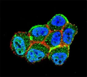
Confocal immunofluorescent analysis of LKB1 antibody (cat # F50221) with ZR-75-1 cells followed by Alexa Fluor 488-conjugated goat anti-rabbit lgG (green). Actin filaments have been labeled with Alexa Fluor 555 Phalloidin (red). DAPI was used as a nuclear counterstain (blue).
|
| ||

|
|
| ||

|