- Tel: 858.663.9055
 Email: info@nsjbio.com
- Tel: 858.663.9055
- Email: info@nsjbio.com
Microglia Marker Antibody detects proteins used to identify microglia, the resident immune cells of the central nervous system (CNS). Microglia arise early in development from yolk sac progenitors and persist throughout life, where they maintain homeostasis, defend against pathogens, and modulate synaptic activity. Unlike peripheral immune cells, microglia remain confined to the brain and spinal cord, adopting dynamic phenotypes in response to environmental cues.
Markers commonly targeted with Microglia Marker Antibodies include Iba1, TMEM119, CD11b, CD68, and CX3CR1. Iba1 is used to visualize microglial morphology, TMEM119 distinguishes resident microglia from infiltrating macrophages, CD68 reflects phagocytosis, and CX3CR1 marks receptor signaling critical for neuron–microglia communication. The Microglia Marker Antibody is therefore essential for accurately identifying microglia in tissue, culture, or disease models.
Because microglial activity underlies both protection and pathology, Microglia Marker Antibodies are central to research in neurodevelopment, neuroinflammation, neurodegeneration, psychiatric disease, and brain injury.
NSJ Bioreagents provides Microglia Marker Antibodies validated for immunohistochemistry, immunofluorescence, flow cytometry, western blotting, and ELISA. Each Microglia Marker Antibody undergoes rigorous testing to confirm specificity for microglial markers, minimizing cross-reactivity with astrocytes, oligodendrocytes, or peripheral immune cells.
By selecting a Microglia Marker Antibody from NSJ Bioreagents, researchers gain reagents optimized for reproducibility. Our antibodies deliver crisp immunostaining in brain tissue, reliable detection in lysates, and dependable performance in multiplex assays. Detailed datasheets, recommended positive controls, and application protocols ensure that Microglia Marker Antibodies support high-quality results across experiments ranging from basic neuroscience to translational medicine.
The Microglia Marker Antibody supports a broad spectrum of applications that reflect the versatile roles of microglia in brain health and disease.
Microglia Marker Antibodies are used to study synaptic pruning during brain development.
The Microglia Marker Antibody highlights microglial contributions to neural circuit formation.
Researchers apply Microglia Marker Antibodies to investigate developmental abnormalities linked to autism spectrum disorders.
The Microglia Marker Antibody is applied in models of stroke, traumatic brain injury, and spinal cord injury.
Microglia Marker Antibodies reveal morphological shifts from surveilling to activated states.
The Microglia Marker Antibody helps clarify microglial involvement in repair versus secondary damage.
In Alzheimer’s disease, Microglia Marker Antibodies detect microglial clustering around amyloid plaques.
The Microglia Marker Antibody supports studies of microglial responses to tau pathology and synaptic loss.
In Parkinson’s, ALS, and Huntington’s disease, Microglia Marker Antibodies highlight inflammatory responses and phagocytic activity.
Microglia Marker Antibodies are applied in depression, schizophrenia, and anxiety research.
The Microglia Marker Antibody helps track how stress alters microglial signaling and behavior.
Microglia Marker Antibodies are used to study viral infections such as HIV, Zika, and herpesvirus in the brain.
The Microglia Marker Antibody supports research into bacterial meningitis and other CNS inflammatory disorders.
Microglia Marker Antibodies validate the effects of drugs designed to suppress harmful microglial activation.
The Microglia Marker Antibody supports biomarker development in cerebrospinal fluid and tissue samples.
In regenerative therapies, Microglia Marker Antibodies evaluate microglial responses to neuronal grafts.
Microglia act as both guardians and potential drivers of pathology in the CNS. The Microglia Marker Antibody allows researchers to monitor these dual roles with precision, while Microglia Marker Antibodies more broadly enable discoveries in neuroscience, pathology, and clinical translation.
In developmental neuroscience, Microglia Marker Antibodies highlight how these cells shape circuits during brain maturation. In neurodegeneration, the Microglia Marker Antibody reveals microglial involvement in Alzheimer’s, Parkinson’s, and ALS. In therapeutics, Microglia Marker Antibodies confirm whether candidate drugs restore balanced microglial activity.
Clinically, microglial activation is increasingly recognized as a biomarker for neurological disorders. The Microglia Marker Antibody links molecular biology with patient care by enabling diagnostic development and treatment monitoring.
Microglia are indispensable regulators of brain health, capable of supporting neural development, mediating defense, or exacerbating disease. The Microglia Marker Antibody equips scientists with a precise and reliable tool for detecting microglia, while Microglia Marker Antibodies more broadly support research in neurodevelopment, neurodegeneration, neuroinflammation, and translational neuroscience. By bridging molecular science with clinical relevance, these antibodies remain vital for advancing neurological health.

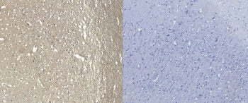
IHC staining of FFPE human frontal cortex tissue with AIF-1 antibody (left) and without AIF-1 antibody (right, negative control).
|
| ||

|
|
| ||

|