- Tel: 858.663.9055
 Email: info@nsjbio.com
- Tel: 858.663.9055
- Email: info@nsjbio.com
Email: info@nsjbio.com
Rabbit monoclonal antibodies combine the specificity of monoclonal antibody technology with the robust immune response naturally generated by rabbits. These reagents have become increasingly popular in research laboratories because they often recognize epitopes that may be difficult to detect using traditional mouse monoclonal antibodies.
Rabbit monoclonal antibodies are widely used in cancer biology, immunology, neuroscience, cardiovascular research, metabolism, and cell biology. Their high affinity and strong target recognition make them valuable tools for detecting proteins expressed at low levels, identifying post-translational modifications, and studying complex signaling pathways.
Rabbit monoclonal antibodies are known for their ability to recognize a broader range of epitopes compared to many traditional mouse monoclonal antibodies. The rabbit immune system frequently generates antibodies against targets that may be weakly immunogenic in other species, allowing researchers to access unique binding specificities.
Because rabbit monoclonal antibodies originate from a single antibody-producing clone, they maintain the consistency and specificity expected from monoclonal reagents while often exhibiting exceptionally strong target affinity. These characteristics can provide improved sensitivity when detecting low-abundance proteins or proteins expressed within challenging tissue environments.
Rabbit monoclonal antibodies are commonly used throughout biomedical research. They are frequently incorporated into studies involving cellular signaling, immune regulation, developmental biology, oncology, and biomarker discovery.
Researchers often utilize rabbit monoclonal antibodies for:
Their versatility makes them valuable tools across a broad range of experimental systems and biological models.
Many cancer-associated biomarkers are routinely detected using rabbit monoclonal antibodies. Their strong affinity and high specificity can support the evaluation of proteins involved in tumor growth, cell proliferation, differentiation, apoptosis, angiogenesis, and immune regulation.
Rabbit monoclonal antibodies are frequently developed against receptors, transcription factors, signaling mediators, and cell surface markers that contribute to tumor progression and therapeutic response. As a result, they have become important tools in oncology research and biomarker development.
Signal transduction pathways depend on precise regulation of protein activation, localization, and expression. Rabbit monoclonal antibodies are commonly used to investigate kinases, phosphoproteins, transcription factors, and intracellular signaling molecules involved in these pathways.
Their ability to recognize highly specific epitopes makes them useful for studying pathway activation, cellular responses to external stimuli, and molecular mechanisms underlying normal physiology and disease.
Because rabbit monoclonal antibodies originate from a single defined clone, they provide a consistent source of target recognition. This clonal origin supports experimental reproducibility and reduces variability that can occur when heterogeneous antibody populations are used.
Modern recombinant production technologies have further improved consistency by enabling controlled expression of defined antibody sequences. These advances help ensure reliable performance across manufacturing lots and long-term supply.
The Rabbit Monoclonal Antibody collection includes antibodies directed against proteins involved in cancer biology, immunology, signal transduction, metabolism, cardiovascular biology, neuroscience, and cell biology. These reagents support investigations into protein expression, cellular signaling, biomarker discovery, and disease-associated molecular mechanisms.
Researchers studying intracellular signaling pathways, receptor activation mechanisms, and cellular regulatory networks may also be interested in our Signal Transduction Antibodies landing page featuring kinases, adaptor proteins, and intracellular signaling related targets.
Browse the complete collection of research antibodies on our Antibodies landing page.

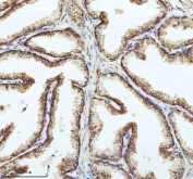
CD1A Antibody Skin Langerhans Cell IHC. Immunohistochemistry analysis of FFPE human skin tissue using CD1A Antibody demonstrates distinct HRP-DAB brown membranous and cytoplasmic staining of epidermal Langerhans cells distributed throughout the epithelial layer. The staining pattern is consistent with the established expression of CD1A on antigen-presenting dendritic cells involved in lipid antigen presentation and immune surveillance at epithelial surfaces. This image highlights the characteristic distribution of CD1A-positive immune cells within the skin and supports studies of dendritic cell biology, antigen presentation, and cutaneous immune responses. Required HIER: boil tissue sections in 10 mM citrate buffer, pH 6, for 10-20 minutes.




 Powered by Bioz




 info@nsjbio.com