- Tel: 858.663.9055
 Email: info@nsjbio.com
- Tel: 858.663.9055
- Email: info@nsjbio.com


| Catalog No | Formulation | Size | Price (USD) | ||
|---|---|---|---|---|---|
 |
R30310 | 0.5mg/ml if reconstituted with 0.2ml sterile DI water | 100 ug | 429 |
| Availability | 1-3 business days |
| Species Reactivity | Human |
| Format | Antigen affinity purified |
| Clonality | Polyclonal (rabbit origin) |
| Isotype | Rabbit IgG |
| Purity | Antigen affinity |
| Buffer | Lyophilized from 1X PBS with 2.5% BSA and 0.025% sodium azide/thimerosal |
| UniProt | P19438 |
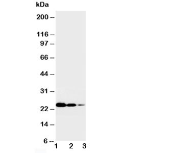
| Applications | Western blot : 0.5-1ug/ml IHC (FFPE) : 0.5-1ug/ml |
| Limitations | This TNFR1 antibody is available for research use only. |
|
Review this product on BioCompare and get a $20 Amazon gift card
|
Related Products
|
Tumor necrosis factor receptor 1(TNFR1), a potent cytokine, elicits a broad spectrum of biologic responses which are mediated by binding to a cell surface receptor. Its gene is located on 12p13.2. The coding region and the 3-prime untranslated region of TNFR1 are distributed over 10 exons. There are 2 different proteins that serve as major receptors for TNF-alpha, one associated with myeloid cells and one associated with epithelial cells. Additionally, TNFR1 associates with the MADD protein through a death domain-death domain interaction. MADD provides a physical link between TNFR1 and the induction of mitogen-activated protein(MAP) kinase(e.g., ERK2) activation and arachidonic acid release. TNFR1-induced apoptosis involves 2 sequential signaling complexes. Complex I, the initial plasma membrane-bound complex, consists of TNFR1, the adaptor TRADD, the kinase RIP1, and TRAF2 and rapidly signals activation of NF-kappa-B. In a second step, TRADD and RIP1 associate with FADD and caspase-8, forming a cytoplasmic complex, complex II.
The stated application concentrations are suggested starting amounts. Titration of the TNFR1 antibody may be required due to differences in protocols and secondary/substrate sensitivity.
An amino acid sequence from the middle region of human TNF Receptor I (CLPQIENVKGTEDSGTT) .
After reconstitution, the TNFR1 antibody can be stored for up to one month at 4oC. For long-term, aliquot and store at -20oC. Avoid repeated freezing and thawing.

Your bulk quote request has been submitted successfully!
Please contact us if you have any questions.
 Powered by Bioz