- Tel: 858.663.9055
 Email: info@nsjbio.com
- Tel: 858.663.9055
- Email: info@nsjbio.com


| Catalog No | Formulation | Size | Price (USD) | ||
|---|---|---|---|---|---|
 |
RQ6741 | 0.5mg/ml if reconstituted with 0.2ml sterile DI water | 100 ug | 429 |
| Availability | 1-3 business days |
| Species Reactivity | Human, Mouse, Rat |
| Format | Purified |
| Clonality | Monoclonal (mouse origin) |
| Isotype | Mouse IgG2a |
| Clone Name | 6G3B4 |
| Purity | Affinity purified |
| Buffer | Lyophilized from 1X PBS with 2% Trehalose |
| UniProt | Q7KZF4 |
| Localization | Cytoplasmic, nuclear |
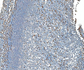
| Applications | Western blot : 1-2ug/ml Immunohistochemistry (FFPE) : 2-5ug/ml Immunofluorescence (FFPE) : 5ug/ml Flow cytometry : 1-3ug/million cells |
| Limitations | This TDRD11 antibody is available for research use only. |
|
Review this product on BioCompare and get a $20 Amazon gift card
|
Related Products
|
Staphylococcal nuclease domain-containing protein 1 also known as 100 kDa coactivator or Tudor domain-containing protein 11 (TDRD11) is a protein that in humans is encoded by the SND1 gene. This gene encodes a transcriptional co-activator that interacts with the acidic domain of Epstein-Barr virus nuclear antigen 2 (EBNA 2), a transcriptional activator that is required for B-lymphocyte transformation. Other transcription factors that interact with this protein are signal transducers and activators of transcription, STATs. This protein is also thought to be essential for normal cell growth. A similar protein in mammals and other organisms is a component of the RNA-induced silencing complex (RISC).
Optimal dilution of the TDRD11 antibody should be determined by the researcher.
Recombinant human protein (amino acids Q20-D204) was used as the immunogen for the TDRD11 antibody.
After reconstitution, the TDRD11 antibody can be stored for up to one month at 4oC. For long-term, aliquot and store at -20oC. Avoid repeated freezing and thawing.

Your bulk quote request has been submitted successfully!
Please contact us if you have any questions.
 Powered by Bioz