- Tel: 858.663.9055
 Email: info@nsjbio.com
- Tel: 858.663.9055
- Email: info@nsjbio.com


| Catalog No | Formulation | Size | Price (USD) | ||
|---|---|---|---|---|---|
 |
RQ6578 | 0.5mg/ml if reconstituted with 0.2ml sterile DI water | 100 ug | 429 |
| Availability | 1-3 business days |
| Species Reactivity | Human, Mouse, Rat |
| Format | Antigen affinity purified |
| Clonality | Polyclonal (rabbit origin) |
| Isotype | Rabbit IgG |
| Purity | Antigen affinity purified |
| Buffer | Lyophilized from 1X PBS with 2% Trehalose |
| UniProt | Q9NYA1 |
| Localization | Nuclear, cytoplasm, cell membrane |
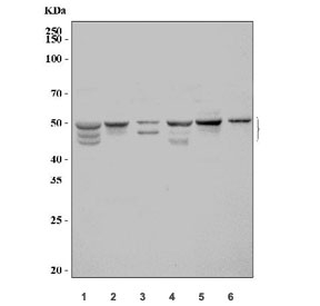
| Applications | Western blot : 1-2ug/ml Immunohistochemistry (FFPE) : 2-5ug/ml Immunofluorescence (FFPE) : 5ug/ml Flow cytometry : 1-3ug/million cells Direct ELISA : 0.1-0.5ug/ml |
| Limitations | This SPHK1 antibody is available for research use only. |
|
Review this product on BioCompare and get a $20 Amazon gift card
|
Related Products
|
SPHK1 (Sphingosine Kinase 1), is an enzyme that in humans is encoded by the SPHK1 gene. Melendez et al.(2000) mapped the SPHK1 gene to chromosome 17q25.2 based on sequence identity with ESTs mapped to this region. Kohama et al.(1998) demonstrated that recombinant mouse Sphk1 can specifically phosphorylate D-erythro-sphingosine and that D, L-threo-dihydrosphingosine and N, N-dimethylsphingosine can act as competitive inhibitors of recombinant Sphk1. Pitson et al.(2000) found that recombinant SPHK1 and endogenous SPHK1 purified from placenta had identical enzymatic characteristics, suggesting posttranslational modification does not effect functional properties.
Optimal dilution of the SPHK1 antibody should be determined by the researcher.
Recombinant human protein (amino acids R16-H267) was used as the immunogen for the SPHK1 antibody.
After reconstitution, the SPHK1 antibody can be stored for up to one month at 4oC. For long-term, aliquot and store at -20oC. Avoid repeated freezing and thawing.

Your bulk quote request has been submitted successfully!
Please contact us if you have any questions.
 Powered by Bioz