- Tel: 858.663.9055
 Email: info@nsjbio.com
- Tel: 858.663.9055
- Email: info@nsjbio.com


| Catalog No | Formulation | Size | Price (USD) | ||
|---|---|---|---|---|---|
 |
V7301-100UG | 0.2 mg/ml in 1X PBS with 0.1 mg/ml BSA (US sourced) and 0.05% sodium azide | 100 ug | 519 | |
|
|
V7301-20UG | 0.2 mg/ml in 1X PBS with 0.1 mg/ml BSA (US sourced) and 0.05% sodium azide | 20 ug | 229 | |
|
|
V7301SAF-100UG | 1 mg/ml in 1X PBS; BSA free, sodium azide free | 100 ug | 519 | |
|
|
V7301IHC-7ML | Prediluted in 1X PBS with 0.1 mg/ml BSA (US sourced) and 0.05% sodium azide; *For IHC use only* | 7 ml | 519 |
| Availability | 1-3 business days |
| Species Reactivity | Human |
| Format | Purified |
| Clonality | Monoclonal (mouse origin) |
| Isotype | Mouse IgG1, kappa |
| Clone Name | RPSA/2699 |
| Purity | Protein G affinity chromatography |
| UniProt | P08865 |
| Localization | Cell surface, cytoplasmic, nuclear |
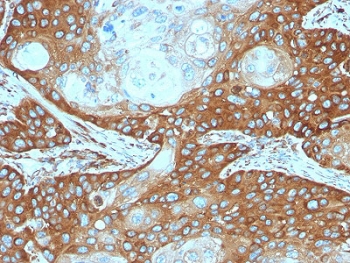
| Applications | Flow cytometry : 1-2ug/ml Immunofluorescence : 1-2ug/ml Western blot : 1-2ug/ml Immunohistochemistry (FFPE) : 1-2ug/ml for 30 min at RT |
| Limitations | This RPSA antibody is available for research use only. |
|
Review this product on BioCompare and get a $20 Amazon gift card
|
Related Products
|
Laminins, a family of extracellular matrix glycoproteins, are the major non-collagenous constituent of basement membranes. They have been implicated in a wide variety of biological processes including cell adhesion, differentiation, migration, signaling, neurite outgrowth and metastasis. Many of the effects of laminin are mediated through interactions with cell surface receptors. These receptors include members of the integrin family, as well as non-integrin laminin-binding proteins. This gene encodes a high-affinity, non-integrin family, laminin receptor 1. Reportedly, level of laminin receptor transcript is higher in colon carcinoma tissue and lung cancer cell line than their normal counterparts. Also, there is a correlation between the upregulation of this polypeptide in cancer cells and their invasive and metastatic phenotype.
Optimal dilution of the RPSA antibody should be determined by the researcher.
1. The prediluted format is supplied in a dropper bottle and is optimized for use in IHC. After epitope retrieval step (if required), drip mAb solution onto the tissue section and incubate at RT for 30 min.
Recombinant full length protein was used as the immunogen for the RPSA antibody.
Store the RPSA antibody at 2-8oC (with azide) or aliquot and store at -20oC or colder (without azide).

Your bulk quote request has been submitted successfully!
Please contact us if you have any questions.

 Powered by Bioz