- Tel: 858.663.9055
 Email: info@nsjbio.com
- Tel: 858.663.9055
- Email: info@nsjbio.com


| Catalog No | Formulation | Size | Price (USD) | ||
|---|---|---|---|---|---|
 |
V7988-100UG | 0.2 mg/ml in 1X PBS with 0.1 mg/ml BSA (US sourced) and 0.05% sodium azide | 100 ug | 519 | |
|
|
V7988-20UG | 0.2 mg/ml in 1X PBS with 0.1 mg/ml BSA (US sourced) and 0.05% sodium azide | 20 ug | 229 | |
|
|
V7988SAF-100UG | 1 mg/ml in 1X PBS; BSA free, sodium azide free | 100 ug | 519 |
| Availability | 1-3 business days |
| Species Reactivity | Human, Mouse |
| Format | Purified |
| Clonality | Monoclonal (mouse origin) |
| Isotype | Mouse IgG1, kappa |
| Clone Name | PDL1/2744 |
| Purity | Protein G affinity chromatography |
| UniProt | Q9NZQ7 |
| Localization | Cell surface, cytoplasmic |
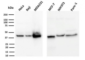
| Applications | ELISA (order BSA-free format for coating) : Western blot : 1-2ug/ml |
| Limitations | This PD-L1 antibody is available for research use only. |
|
Review this product on BioCompare and get a $20 Amazon gift card
|
Related Products
|
Engagement of CD28 by B7-1 (CD80) or B7-2 (CD86) in the presence of antigen promotes T-cell proliferation, cytokine production, differentiation of effector T-cells and the induction of BCLX, a promoter of T-cell survival. Engagement of CTLA4 by B7-1 or B7-2, on the other hand, may inhibit proliferation and interleukin-2 (IL-2) production. PD-L1 is 290-amino acid type I transmembrane protein, which is 20% and 15% identical to B7-1 and B7-2, respectively, has immunoglobulin V-like and C-like domains and a 30-amino acid cytoplasmic tail. PD-L1 does not bind CD28, cytotoxic T-lymphocyte A4 or ICOS (inducible co-stimulator). IL-2, although produced in small amounts, is required for the effect of PD-L1 co-stimulation. PD-L2 protein contains a signal sequence, IgV- and IgC-like domains, a transmembrane region and a cytoplasmic region. The constitutive expression of PD-L1 and PD-L2 on parenchymal cells of heart, lung and kidney suggests that the PD-1-PD-L system could provide unique negative signaling to help prevent autoimmune diseases.
Optimal dilution of the PD-L1 antibody should be determined by the researcher.
A recombinant human partial protein (amino acids 39-191) was used as the immunogen for the PD-L1 antibody.
Store the PD-L1 antibody at 2-8oC (with azide) or aliquot and store at -20oC or colder (without azide).

Your bulk quote request has been submitted successfully!
Please contact us if you have any questions.

 Powered by Bioz