- Tel: 858.663.9055
 Email: info@nsjbio.com
- Tel: 858.663.9055
- Email: info@nsjbio.com


| Catalog No | Formulation | Size | Price (USD) | ||
|---|---|---|---|---|---|
 |
R30902 | 0.5mg/ml if reconstituted with 0.2ml sterile DI water | 100 ug | 429 |
| Availability | 1-3 business days |
| Species Reactivity | Human, Mouse, Rat |
| Format | Antigen affinity purified |
| Clonality | Polyclonal (rabbit origin) |
| Isotype | Rabbit IgG |
| Purity | Antigen affinity |
| Buffer | Lyophilized from 1X PBS with 2.5% BSA and 0.025% sodium azide/thimerosal |
| UniProt | P49023 |
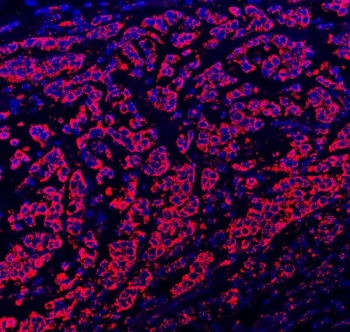
| Applications | Western blot : 0.5-1ug/ml Immunohistochemistry (FFPE) : 0.5-1ug/ml Iimmunohistochemistry (Frozen) : 0.5-1ug/ml Immunocytochemistry : 0.5-1ug/ml Immunofluorescence : 5ug/ml Flow cytometry : 1-3ug/million cells |
| Limitations | This Paxillin antibody is available for research use only. |
|
Review this product on BioCompare and get a $20 Amazon gift card
|
Related Products
|
Paxillin is a signal transduction adaptor protein discovered in 1990 in the laboratory of Keith Burridge. Salgia et al.(1995) mapped the gene to 12q24 using fluorescence in situ hybridization.The C-terminal region of Paxillin contains four LIM domains that target paxillin to focal adhesions, it is presumed through a direct association with the cytoplasmic tail of beta-integrin. The N-terminal region is rich in protein-protein interaction sites. The proteins that bind to Paxillin are diverse and include protein tyrosine kinases, such as Src and FAK, structural proteins, such as vinculin and actopaxin, and regulators of actin organization, such as COOL/PIX and PKL/GIT. Paxillin is tyrosine-phosphorylated by FAK and Src upon integrin engagement or growth factor stimulation, creating binding sites for the adapter protein Crk. The protein contains 4 LIM domains, a proline-rich domain containing a consensus SH3-binding site, and 3 potential SH2-binding sites.
The stated application concentrations are suggested starting amounts. Titration of the Paxillin antibody may be required due to differences in protocols and secondary/substrate sensitivity.
Amino acids 456-472 (HEKDGKAYCRKDYFDMF) were used as the immunogen for this Paxillin antibody (100% homologous in human, mouse and rat).
After reconstitution, the Paxillin antibody can be stored for up to one month at 4oC. For long-term, aliquot and store at -20oC. Avoid repeated freezing and thawing.

Your bulk quote request has been submitted successfully!
Please contact us if you have any questions.
 Powered by Bioz