- Tel: 858.663.9055
 Email: info@nsjbio.com
- Tel: 858.663.9055
- Email: info@nsjbio.com


| Catalog No | Formulation | Size | Price (USD) | ||
|---|---|---|---|---|---|
 |
V3168-100UG | 0.2 mg/ml in 1X PBS with 0.1 mg/ml BSA (US sourced) and 0.05% sodium azide | 100 ug | 519 | |
|
|
V3168-20UG | 0.2 mg/ml in 1X PBS with 0.1 mg/ml BSA (US sourced) and 0.05% sodium azide | 20 ug | 199 | |
|
|
V3168SAF-100UG | 1 mg/ml in 1X PBS; BSA free, sodium azide free | 100 ug | 429 | |
|
|
V3168IHC-7ML | Prediluted in 1X PBS with 0.1 mg/ml BSA (US sourced) and 0.05% sodium azide; *For IHC use only* | 7 ml | 429 |
| Availability | 1-3 business days |
| Species Reactivity | Human, Mouse, Rat. Other species not known. |
| Format | Purified |
| Clonality | Monoclonal (mouse origin) |
| Isotype | Mouse IgG1, kappa |
| Clone Name | 5.8A |
| Purity | Protein G affinity chromatography |
| UniProt | P15172 |
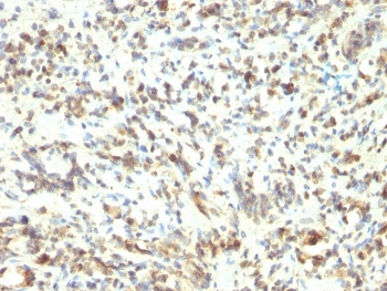
| Localization | Nuclear. Only nuclear staining should be considered as evidence of skeletal muscle differentiation. |
| Applications | Flow Cytometry : 0.5-1ug/million cells in 0.1ml Immunofluorescence : 0.5-1ug/ml Immunohistochemistry (FFPE) : 0.5-1ug/ml for 30 min at RT (1) Prediluted IHC only format : incubate for 30 min at RT (2) |
| Limitations | This MyoD1 antibody is available for research use only. |
|
Review this product on BioCompare and get a $20 Amazon gift card
|
Related Products
|
Recognizes a phosphor-protein of 45kDa, identified as MyoD1. It does not cross react with myogenin, Myf5, or Myf6. Antibody to MyoD1 labels the nuclei of myoblasts in developing muscle tissues. MyoD1 is not detected in normal adult tissue, but is highly expressed in the tumor cell nuclei of rhabdomyosarcomas. Occasionally nuclear expression of MyoD1 is seen in ectomesenchymoma and a subset of Wilms tumors. Weak cytoplasmic staining is observed in several non-muscle tissues, including glandular epithelium and also in rhabdomyosarcomas, neuroblastomas, Ewing s sarcomas and alveolar soft part sarcomas.
The optimal dilution of the MyoD1 antibody for each application should be determined by the researcher.
1. The prediluted format is supplied in a dropper bottle and is optimized for use in IHC. After epitope retrieval step (if required), drip mAb solution onto the tissue section and incubate at RT for 30 min.
Recombinant mouse MyoD1 protein was used as the immunogen for this MyoD1 antibody. The epitope of this mAb maps between amino acid 180-189 in the C-terminal of mouse MyoD1 protein.
Store the MyoD1 antibody at 2-8oC (with azide) or aliquot and store at -20oC or colder (without azide).

Your bulk quote request has been submitted successfully!
Please contact us if you have any questions.
 Powered by Bioz