- Tel: 858.663.9055
 Email: info@nsjbio.com
- Tel: 858.663.9055
- Email: info@nsjbio.com


| Catalog No | Formulation | Size | Price (USD) | ||
|---|---|---|---|---|---|
 |
RQ6388 | 0.5mg/ml if reconstituted with 0.2ml sterile DI water | 100 ug | 429 |
| Availability | 1-3 business days |
| Species Reactivity | Human |
| Format | Purified |
| Clonality | Monoclonal (mouse origin) |
| Isotype | Mouse IgG2b |
| Clone Name | 9B6 |
| Purity | Antigen affinity purified |
| Buffer | Lyophilized from 1X PBS with 2% Trehalose |
| UniProt | P12956 |
| Localization | Nuclear |
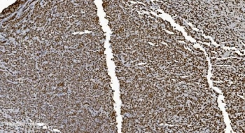
| Applications | Western blot : 0.5-1ug/ml Immunohistochemistry (FFPE) : 2-5ug/ml Immunofluorescence (FFPE) : 5ug/ml Flow cytometry : 1-3ug/million cells |
| Limitations | This Ku70 antibody is available for research use only. |
|
Review this product on BioCompare and get a $20 Amazon gift card
|
Related Products
|
XRCC6 (X-Ray Repair, Complementing Defective, In Chinese Hamster, 6), also called Ku70, G22P1 or TLAA, is a protein that in humans, is encoded by the XRCC6 gene. In addition, the XRCC6 gene encodes subunit p70 of the p70/p80 autoantigen which consists of 2 proteins of molecular mass of approximately 70,000 and 80,000 daltons that dimerize to form a 10 S DNA-binding complex. The XRCC6 gene is mapped to 22q13.2. XRCC6 and Mre11 are differentially expressed during meiosis. XRCC6 interacts with Baxa, a mediator of mitochondrial-dependent apoptosis. Disruption of both FANCC and XRCC6 suppressed sensitivity to crosslinking agents, diminished chromosome breaks, and reversed defective homologous recombination. Ku70 binds directly to free DNA ends, committing them to NHEJ repair. In early meiotic prophase, however, when meiotic recombination is most probably initiated, Mre11 was abundant, whereas XRCC6 was not detectable.
Optimal dilution of the Ku70 antibody should be determined by the researcher.
Amino acids AIVEKLRFTYRSDSFENPVLQQHFRNLEALALD were used as the immunogen for the Ku70 antibody.
After reconstitution, the Ku70 antibody can be stored for up to one month at 4oC. For long-term, aliquot and store at -20oC. Avoid repeated freezing and thawing.

Your bulk quote request has been submitted successfully!
Please contact us if you have any questions.
 Powered by Bioz