- Tel: 858.663.9055
 Email: info@nsjbio.com
- Tel: 858.663.9055
- Email: info@nsjbio.com


| Catalog No | Formulation | Size | Price (USD) | ||
|---|---|---|---|---|---|
 |
V2604-100UG | 0.2 mg/ml in 1X PBS with 0.1 mg/ml BSA (US sourced) and 0.05% sodium azide | 100 ug | 519 | |
|
|
V2604-20UG | 0.2 mg/ml in 1X PBS with 0.1 mg/ml BSA (US sourced) and 0.05% sodium azide | 20 ug | 229 | |
|
|
V2604SAF-100UG | 1 mg/ml in 1X PBS; BSA free, sodium azide free | 100 ug | 519 | |
|
|
V2604IHC-7ML | Prediluted in 1X PBS with 0.1 mg/ml BSA (US sourced) and 0.05% sodium azide; *For IHC use only* | 7 ml | 519 |
| Availability | 1-3 business days |
| Species Reactivity | Human. Other species not known. |
| Format | Purified |
| Clonality | Monoclonal (mouse origin) |
| Isotype | Mouse IgG2b, kappa |
| Clone Name | 1H4 or W-CAM-1 or Wehi-CAM-1 |
| Purity | Protein G affinity chromatography |
| UniProt | P05362 |
| Localization | Cell surface |
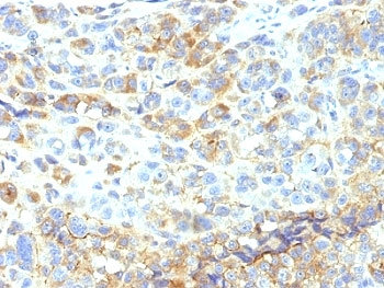
| Applications | Flow cytometry : 0.5-1ug/million cells in 0.1ml Functional testing (order BSA/sodium azide-free format) : Immunohistochemistry (FFPE) : 2-4ug/ml for 30 min at RT (1) Prediluted IHC only format : incubate for 30 min at RT (2) |
| Limitations | This ICAM-1 antibody is available for research use only. |
|
Review this product on BioCompare and get a $20 Amazon gift card
|
Related Products
|
Recognizes an 85-115kDa protein (variation with cell type), identified as intercellular adhesion molecule (ICAM-1) (Workshop IV). It has 7 potential N-linked glycosylation sites. ICAM-1 is a single chain glycoprotein of Ig supergene family, present on unstimulated endothelial cells (EC) and on a variety of other cell types including activated fibroblasts, EC, macrophages, and lymphocytes. ICAM-1 mediates cell adhesion by binding to integrins CD11a/CD18 (leukocyte adhesion molecule, LFA-1) and to CD11b/CD18 (Mac-1). This interaction enhances antigen-specific T-cell activation. ICAM-1 also binds to CD43 and to Plasmodium falciparum infected RBCs. W-CAM-1 mAb blocks aggregation of cell lines mediated by the ICAM-1 and blocks homotypic binding of purified populations of activated T- and B-lymphocytes and also aggregation of mixed T- and B-cell blasts. It inhibits T-cell adhesion to normal human endothelial cells. Activation induced by cell-cell contact (mixed lymphocyte reaction, T-cell mediated B-cell activation) is significantly inhibited. This mAb blocks elements of both effector arms of immune system (cytotoxic cell function and Ig production).
Optimal dilution of the ICAM-1 antibody should be determined by the researcher.
1. Staining of formalin-fixed tissues requires boiling tissue sections in 10mM Tris with 1mM EDTA, pH 9.0 for 10-20 min followed by cooling at RT for 20 min
2. The prediluted format is supplied in a dropper bottle and is optimized for use in IHC. After epitope retrieval step (if required), drip mAb solution onto the tissue section and incubate at RT for 30 min.
Raji Burkitt lymphoma cells were used as the immunogen for the ICAM-1 antibody.
Store the ICAM-1 antibody at 2-8oC (with azide) or aliquot and store at -20oC or colder (without azide).

Your bulk quote request has been submitted successfully!
Please contact us if you have any questions.
 Powered by Bioz