- Tel: 858.663.9055
 Email: info@nsjbio.com
- Tel: 858.663.9055
- Email: info@nsjbio.com


| Catalog No | Formulation | Size | Price (USD) | ||
|---|---|---|---|---|---|
 |
V2566-100UG | 0.2 mg/ml in 1X PBS with 0.1 mg/ml BSA (US sourced) and 0.05% sodium azide | 100 ug | 519 | |
|
|
V2566-20UG | 0.2 mg/ml in 1X PBS with 0.1 mg/ml BSA (US sourced) and 0.05% sodium azide | 20 ug | 229 | |
|
|
V2566SAF-100UG | 1 mg/ml in 1X PBS; BSA free, sodium azide free | 100 ug | 519 | |
|
|
V2566IHC-7ML | Prediluted in 1X PBS with 0.1 mg/ml BSA (US sourced) and 0.05% sodium azide; *For IHC use only* | 7 ml | 519 |
| Availability | 1-3 business days |
| Species Reactivity | Human, Mouse, Rat |
| Format | Purified |
| Clonality | Monoclonal (mouse origin) |
| Isotype | Mouse IgG2a, kappa |
| Clone Name | SPM256 |
| Purity | Protein G affinity chromatography |
| UniProt | P07305 |
| Localization | Nuclear |
| Applications | Immunofluorescence : 1-2ug/ml Immunohistochemistry (FFPE) : 1-2ug/ml for 30 min at RT Western blot : 1-2ug/ml Flow cytometry : 1-2ug/10^6 cells |
| Limitations | This anti-Histone H1 antibody is available for research use only. |
|
Review this product on BioCompare and get a $20 Amazon gift card
|
Related Products
|
Eukaryotic histones are basic and water-soluble nuclear proteins that form hetero-octameric nucleosome particles by wrapping 146 base pairs of DNA in a left-handed super-helical turn sequentially to form chromosomal fiber. Two molecules of each of the four core histones (H2A, H2B, H3, and H4) form the octamer; formed of two H2A-H2B dimers and two H3-H4 dimers, forming two nearly symmetrical halves by tertiary structure. Over 80% of nucleosomes contain the linker Histone H1, derived from an intronless gene that interacts with linker DNA between nucleosomes and mediates compaction into higher order chromatin. Histones are subject to posttranslational modification by enzymes primarily on their N-terminal tails, but also in their globular domains. Such modifications include methylation, citrullination, acetylation, phosphorylation, sumoylation, ubiquitination and ADP-ribosylation.
Optimal dilution of the anti-Histone H1 antibody should be determined by the researcher.
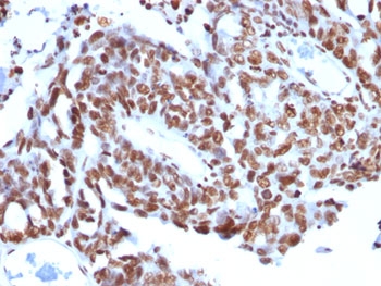
1. Staining of formalin/paraffin tissues requires boiling tissue sections in pH 9 10mM Tris with 1mM EDTA for 10-20 min followed by cooling at RT for 20 min.
2. The prediluted format is supplied in a dropper bottle and is optimized for use in IHC. After epitope retrieval step (if required), drip mAb solution onto the tissue section and incubate at RT for 30 min.
Nuclei of human leukemia biopsy cells were used as the immunogen for the anti-Histone H1 antibody.
Store the anti-Histone H1 antibody at 2-8oC (with azide) or aliquot and store at -20oC or colder (without azide).

Your bulk quote request has been submitted successfully!
Please contact us if you have any questions.
 Powered by Bioz