- Tel: 858.663.9055
 Email: info@nsjbio.com
- Tel: 858.663.9055
- Email: info@nsjbio.com


| Catalog No | Formulation | Size | Price (USD) | ||
|---|---|---|---|---|---|
 |
RQ6424 | 0.5mg/ml if reconstituted with 0.2ml sterile DI water | 100 ug | 429 |
| Availability | 1-3 business days |
| Species Reactivity | Human, Mouse, Rat |
| Format | Purified |
| Clonality | Polyclonal (rabbit origin) |
| Isotype | Rabbit IgG |
| Purity | Antigen affinity purified |
| Buffer | Lyophilized from 1X PBS with 2% Trehalose |
| UniProt | P00505 |
| Localization | Cytoplasmic, cell membrane |
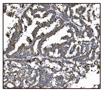
| Applications | Western blot : 0.5-1ug/ml Immunohistochemistry (FFPE) : 2-5ug/ml Flow cytometry : 1-3ug/million cells Direct ELISA : 0.1-0.5ug/ml |
| Limitations | This Glutamate-oxaloacetate transaminase 2 antibody is available for research use only. |
|
Review this product on BioCompare and get a $20 Amazon gift card
|
Related Products
|
Aspartate aminotransferase, mitochondrial is an enzyme that in humans is encoded by the GOT2 gene. Glutamic-oxaloacetic transaminase is a pyridoxal phosphate-dependent enzyme which exists in cytoplasmic and inner-membrane mitochondrial forms, GOT1 and GOT2, respectively. GOT plays a role in amino acid metabolism and the urea and tricarboxylic acid cycles. The two enzymes are homodimeric and show close homology. Two transcript variants encoding different isoforms have been found for this gene.
Optimal dilution of the Glutamate-oxaloacetate transaminase 2 antibody should be determined by the researcher.
Recombinant human protein (amino acids H35-K430) was used as the immunogen for the Glutamate-oxaloacetate transaminase 2 antibody.
After reconstitution, the Glutamate-oxaloacetate transaminase 2 antibody can be stored for up to one month at 4oC. For long-term, aliquot and store at -20oC. Avoid repeated freezing and thawing.

Your bulk quote request has been submitted successfully!
Please contact us if you have any questions.
 Powered by Bioz