- Tel: 858.663.9055
 Email: info@nsjbio.com
- Tel: 858.663.9055
- Email: info@nsjbio.com

| Catalog No | Formulation | Size | Price (USD) | ||
|---|---|---|---|---|---|
 |
RQ6231 | 0.5mg/ml if reconstituted with 0.2ml sterile DI water | 100 ug | 429 |
| Availability | 1-3 business days |
| Species Reactivity | Human, Mouse, Rat |
| Format | Antigen affinity purified |
| Clonality | Monoclonal (mouse origin) |
| Isotype | Mouse IgG2a |
| Clone Name | 3B6 |
| Purity | Affinity purified |
| Buffer | Lyophilized from 1X PBS with 2% Trehalose |
| UniProt | P62495 |
| Localization | Cytoplasmic |
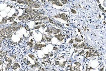
| Applications | Western blot : 1-2ug/ml Immunohistochemistry (FFPE) : 2-5ug/ml Immunofluorescence : 5ug/ml Flow cytometry : 1-3ug/million cells |
| Limitations | This eRF1 antibody is available for research use only. |
|
Review this product on BioCompare and get a $20 Amazon gift card
|
Related Products
|
Eukaryotic translation termination factor 1 (eRF1), also known as TB3-1, is a protein that in humans is encoded by the ETF1 gene. It is mapped to 5q31.2. This gene encodes a class-1 polypeptide chain release factor. The encoded protein plays an essential role in directing termination of mRNA translation from the termination codons UAA, UAG and UGA. This protein is a component of the SURF complex which promotes degradation of prematurely terminated mRNAs via the mechanism of nonsense-mediated mRNA decay (NMD). Alternate splicing results in multiple transcript variants. Pseudogenes of this gene are found on chromosomes 6, 7, and X.
Optimal dilution of the eRF1 antibody should be determined by the researcher.
A human recombinant partial protein (amino acids D9-K342) was used as the immunogen for the eRF1 antibody.
After reconstitution, the eRF1 antibody can be stored for up to one month at 4oC. For long-term, aliquot and store at -20oC. Avoid repeated freezing and thawing.

Your bulk quote request has been submitted successfully!
Please contact us if you have any questions.
 Powered by Bioz