- Tel: 858.663.9055
 Email: info@nsjbio.com
- Tel: 858.663.9055
- Email: info@nsjbio.com


| Catalog No | Formulation | Size | Price (USD) | ||
|---|---|---|---|---|---|
 |
V3053-100UG | 0.2 mg/ml in 1X PBS with 0.1 mg/ml BSA (US sourced) and 0.05% sodium azide | 100 ug | 519 | |
|
|
V3053-20UG | 0.2 mg/ml in 1X PBS with 0.1 mg/ml BSA (US sourced) and 0.05% sodium azide | 20 ug | 229 | |
|
|
V3053SAF-100UG | 1 mg/ml in 1X PBS; BSA free, sodium azide free | 100 ug | 519 | |
|
|
V3053IHC-7ML | Prediluted in 1X PBS with 0.1 mg/ml BSA (US sourced) and 0.05% sodium azide; *For IHC use only* | 7 ml | 519 |
| Availability | 1-3 business days |
| Species Reactivity | Human |
| Format | Purified |
| Clonality | Monoclonal (mouse origin) |
| Isotype | Mouse IgG2a, kappa |
| Clone Name | DE-K13 |
| Purity | Protein G affinity chromatography |
| UniProt | P13645 , P13646 |
| Localization | Cytoplasmic |
| Applications | Flow cytometry : 1-2ug/10^6 cells Immunofluorescence : 1-2ug/ml Western blot : 1-2ug/ml Immunohistochemistry (FFPE) : 1-2ug/ml for 30 min at RT |
| Limitations | This Cytokeratin 10/13 antibody is available for research use only. |
|
Review this product on BioCompare and get a $20 Amazon gift card
|
Related Products
|
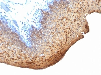
This antibody recognizes cytokeratin 10 (56.5kDa) and cytokeratin 13 (53kDa) in Western blotting. It recognizes only cytokeratin 13 in formalin-fixed, paraffin-embedded tissue sections. It does not react with cytokeratin 10 positive, cytokeratin 13 negative epithelia such as epidermis. However, on tissue sections this mAb serves as differentiation-related marker of all stratified epithelia; it stains all suprabasal cells in both cornifying and non-cornifying stratified epithelia and more differentiated cells of squamous carcinomas.
Optimal dilution of the Cytokeratin 10/13 antibody should be determined by the researcher.
1. Staining of formalin-fixed tissues requires boiling tissue sections in 10mM Citrate buffer, pH 6.0, for 10-20 min followed by cooling at RT for 20 min.
2. The prediluted format is supplied in a dropper bottle and is optimized for use in IHC. After epitope retrieval step (if required), drip mAb solution onto the tissue section and incubate at RT for 30 min.
A cytoskeletal preparation extracted from human ectocervical epithelium was used as the immunogen for the Cytokeratin 10/13 antibody.
Store the Cytokeratin 10/13 antibody at 2-8oC (with azide) or aliquot and store at -20oC or colder (without azide).

Your bulk quote request has been submitted successfully!
Please contact us if you have any questions.
 Powered by Bioz