- Tel: 858.663.9055
 Email: info@nsjbio.com
- Tel: 858.663.9055
- Email: info@nsjbio.com


| Catalog No | Formulation | Size | Price (USD) | ||
|---|---|---|---|---|---|
 |
V2915-100UG | 0.2 mg/ml in 1X PBS with 0.1 mg/ml BSA (US sourced) and 0.05% sodium azide | 100 ug | 519 | |
|
|
V2915-20UG | 0.2 mg/ml in 1X PBS with 0.1 mg/ml BSA (US sourced) and 0.05% sodium azide | 20 ug | 229 | |
|
|
V2915SAF-100UG | 1 mg/ml in 1X PBS; BSA free, sodium azide free | 100 ug | 519 | |
|
|
V2915IHC-7ML | Prediluted in 1X PBS with 0.1 mg/ml BSA (US sourced) and 0.05% sodium azide; *For IHC use only* | 7 ml | 519 |
| Availability | 1-3 business days |
| Species Reactivity | Human |
| Format | Purified |
| Clonality | Monoclonal (mouse origin) |
| Isotype | Mouse IgG2a, kappa |
| Clone Name | T311 + OCA1/812 |
| Purity | Protein G affinity chromatography |
| UniProt | P14679 |
| Localization | Cytoplasmic |
| Applications | Immunohistochemistry (FFPE) : 1-2ug/ml for 30 min at RT |
| Limitations | This TYR antibody cocktail is available for research use only. |
|
Review this product on BioCompare and get a $20 Amazon gift card
|
Related Products
|
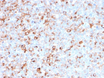
Recognizes a cluster of proteins between 70-80kDa, identified as Tyrosinase/TYR. Occasionally a minor band at 55kDa is also detected. This mAb shows no cross-reaction with MAGE-1 and tyrosinase-related protein 1, TRP-1/gp75. Tyrosinase is a copper-containing metalloglycoprotein that catalyzes several steps in the melanin pigment biosynthetic pathway; the hydroxylation of tyrosine to L-3,4-dihydroxy-phenylalanine (dopa), and the subsequent oxidation of dopa to dopaquinone. Mutations of the tyrosinase gene occur in various forms of albinism. Tyrosinase is one of the targets for cytotoxic T-cell recognition in melanoma patients. Staining of melanomas with this mAb shows tyrosinase in melanotic as well as amelanotic variants. This mAb is a useful marker for melanocytes and melanomas.
Optimal dilution of the TYR antibody cocktail should be determined by the researcher.
1. Staining of formalin-fixed tissues requires boiling tissue sections in 10mM Tris with 1mM EDTA, pH 9, for 10-20 min followed by cooling at RT for 20 min.
2. The prediluted format is supplied in a dropper bottle and is optimized for use in IHC. After epitope retrieval step (if required), drip mAb solution onto the tissue section and incubate at RT for 30 min.
Recombinant human protein was used as the immunogen for the TYR antibody cocktail.
Store the TYR antibody cocktail at 2-8oC (with azide) or aliquot and store at -20oC or colder (without azide).

Your bulk quote request has been submitted successfully!
Please contact us if you have any questions.
 Powered by Bioz