- Tel: 858.663.9055
 Email: info@nsjbio.com
- Tel: 858.663.9055
- Email: info@nsjbio.com

| Catalog No | Formulation | Size | Price (USD) | ||
|---|---|---|---|---|---|
 |
RQ6290 | 0.5mg/ml if reconstituted with 0.2ml sterile DI water | 100 ug | 429 |
| Availability | 1-3 business days |
| Species Reactivity | Human, Mouse, Rat |
| Format | Antigen affinity purified |
| Clonality | Monoclonal (mouse origin) |
| Isotype | Mouse IgG2b |
| Clone Name | 7B12 |
| Purity | Affinity purified |
| Buffer | Lyophilized from 1X PBS with 2% Trehalose |
| UniProt | Q71U36/P68363/Q9BQE3 |
| Localization | Cytoplasmic |
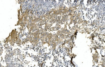
| Applications | Western blot : 1-2ug/ml Immunohistochemistry (FFPE) : 2-5ug/ml Immunofluorescence : 5ug/ml Flow cytometry : 1-3ug/million cells |
| Limitations | This TUBA1 antibody is available for research use only. |
|
Review this product on BioCompare and get a $20 Amazon gift card
|
Related Products
|
Tubulin is the major constituent of microtubules. Microtubules of the eukaryotic cytoskeleton perform essential and diverse functions and are composed of a heterodimer of alpha and beta tubulins.
Optimal dilution of the TUBA1 antibody should be determined by the researcher.
A human recombinant partial protein (amino acids N18-A403) was used as the immunogen for the TUBA1 antibody.
After reconstitution, the TUBA1 antibody can be stored for up to one month at 4oC. For long-term, aliquot and store at -20oC. Avoid repeated freezing and thawing.

Your bulk quote request has been submitted successfully!
Please contact us if you have any questions.
 Powered by Bioz