- Tel: 858.663.9055
 Email: info@nsjbio.com
- Tel: 858.663.9055
- Email: info@nsjbio.com


| Catalog No | Formulation | Size | Price (USD) | ||
|---|---|---|---|---|---|
 |
RQ7543 | 0.5mg/ml if reconstituted with 0.2ml sterile DI water | 100 ug | 429 |
| Availability | 1-3 business days |
| Species Reactivity | Human, Mouse, Rat |
| Format | Antigen affinity purified |
| Clonality | Polyclonal (rabbit origin) |
| Isotype | Rabbit IgG |
| Purity | Antigen affinity purified |
| Buffer | Lyophilized from 1X PBS with 2% Trehalose |
| UniProt | O15050 |
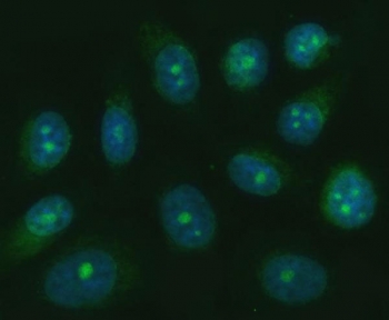
| Applications | Western blot : 0.5-1ug/ml Immunohistochemistry (FFPE) : 2-5ug/ml Immunofluorescence : 5ug/ml Flow cytometry : 1-3ug/million cells Direct ELISA : 0.1-0.5ug/ml |
| Limitations | This TRANK1 antibody is available for research use only. |
|
Review this product on BioCompare and get a $20 Amazon gift card
|
Tetratricopeptide repeat and ankyrin repeat containing 1 is a protein that in humans is encoded by the TRANK1 gene. Predicted to enable ATP binding activity.
Optimal dilution of the TRANK1 antibody should be determined by the researcher.
E. coli-derived recombinant human protein (amino acids K1926-R2763) was used as the immunogen for the TRANK1 antibody.
After reconstitution, the TRANK1 antibody can be stored for up to one month at 4oC. For long-term, aliquot and store at -20oC. Avoid repeated freezing and thawing.

Your bulk quote request has been submitted successfully!
Please contact us if you have any questions.
 Powered by Bioz