- Tel: 858.663.9055
 Email: info@nsjbio.com
- Tel: 858.663.9055
- Email: info@nsjbio.com


| Catalog No | Formulation | Size | Price (USD) | ||
|---|---|---|---|---|---|
 |
RQ4532 | 0.5mg/ml if reconstituted with 0.2ml sterile DI water | 100 ug | 429 |
| Availability | 1-3 business days |
| Species Reactivity | Human, Mouse, Rat |
| Format | Antigen affinity purified |
| Clonality | Polyclonal (rabbit origin) |
| Isotype | Rabbit IgG |
| Purity | Antigen affinity |
| Buffer | Lyophilized from 1X PBS with 2% Trehalose and 0.025% sodium azide |
| UniProt | P08047 |
| Localization | Nuclear, cytoplasmic |
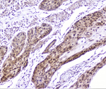
| Applications | Western blot : 0.1-0.5ug/ml IHC (FFPE) : 1-2ug/ml Immunofluorescence/Immunocytochemistry : 2-4ug/ml Immunocytochemistry : 1-2ug/ml Direct ELISA : 0.1-0.5ug/ml (human recombinant protein) |
| Limitations | This SP1 antibody is available for research use only. |
|
Review this product on BioCompare and get a $20 Amazon gift card
|
Related Products
|
Transcription factor Sp1, also known as 'Specificity protein 1' is a protein that in humans is encoded by the SP1 gene. The protein encoded by this gene is a zinc finger transcription factor that binds to GC-rich motifs of many promoters. The encoded protein is involved in many cellular processes, including cell differentiation, cell growth, apoptosis, immune responses, response to DNA damage, and chromatin remodeling. Post-translational modifications such as phosphorylation, acetylation, glycosylation, and proteolytic processing significantly affect the activity of this protein, which can be an activator or a repressor. Three transcript variants encoding different isoforms have been found for this gene.
Optimal dilution of the SP1 antibody should be determined by the researcher.
Amino acids 384-603 of the human protein were used as the immunogen for the SP1 antibody.
After reconstitution, the SP1 antibody can be stored for up to one month at 4oC. For long-term, aliquot and store at -20oC. Avoid repeated freezing and thawing.

Your bulk quote request has been submitted successfully!
Please contact us if you have any questions.
 Powered by Bioz