- Tel: 858.663.9055
 Email: info@nsjbio.com
- Tel: 858.663.9055
- Email: info@nsjbio.com


| Catalog No | Formulation | Size | Price (USD) | ||
|---|---|---|---|---|---|
 |
RQ7022 | 0.5mg/ml if reconstituted with 0.2ml sterile DI water | 100 ug | 429 |
| Availability | 1-3 business days |
| Species Reactivity | Human, Monkey |
| Format | Antigen affinity purified |
| Clonality | Monoclonal (mouse origin) |
| Isotype | Mouse IgG2b |
| Clone Name | 3H5E7 |
| Purity | Antigen affinity purified |
| Buffer | Lyophilized from 1X PBS with 2% Trehalose |
| UniProt | Q15084 |
| Localization | Cytoplasmic, cell membrane |
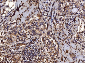
| Applications | Western blot : 0.5-1 ug/ml Immunohistochemistry (FFPE) : 2-5ug/ml Immunofluorescence : 5ug/ml Flow cytometry : 1-3ug/million cells |
| Limitations | This PDIA6 antibody is available for research use only. |
|
Review this product on BioCompare and get a $20 Amazon gift card
|
Related Products
|
This gene encodes a member of the disulfide isomerase (PDI) family of endoplasmic reticulum (ER) proteins that catalyze protein folding and thiol-disulfide interchange reactions. The encoded protein has an N-terminal ER-signal sequence, two catalytically active thioredoxin (TRX) domains, a TRX-like domain, and a C-terminal ER-retention sequence. This protein inhibits the aggregation of misfolded proteins and exhibits both isomerase and chaperone activity. Alternative splicing results in multiple transcript variants encoding different isoforms.
Optimal dilution of the PDIA6 antibody should be determined by the researcher.
Recombinant human protein (amino acids L20-L440) was used as the immunogen for the PDIA6 antibody.
After reconstitution, the PDIA6 antibody can be stored for up to one month at 4oC. For long-term, aliquot and store at -20oC. Avoid repeated freezing and thawing.

Your bulk quote request has been submitted successfully!
Please contact us if you have any questions.
 Powered by Bioz