- Tel: 858.663.9055
 Email: info@nsjbio.com
- Tel: 858.663.9055
- Email: info@nsjbio.com


| Catalog No | Formulation | Size | Price (USD) | ||
|---|---|---|---|---|---|
 |
R31058 | 0.5mg/ml if reconstituted with 0.2ml sterile DI water | 100 ug | 429 |
| Availability | 1-3 business days |
| Species Reactivity | Human, Mouse, Rat |
| Format | Antigen affinity purified |
| Clonality | Polyclonal (rabbit origin) |
| Isotype | Rabbit IgG |
| Purity | Antigen affinity |
| Buffer | Lyophilized from 1X PBS with 2.5% BSA and 0.025% sodium azide/thimerosal |
| UniProt | Q13501 |
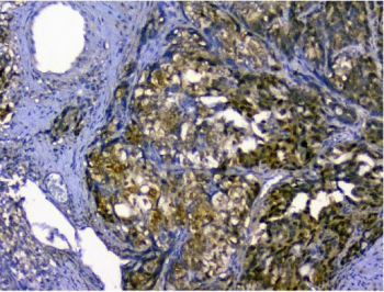
| Applications | Western blot : 0.5-1ug/ml Immunohistochemistry (FFPE) : 0.5-1ug/ml Immunocytochemistry : 0.5-1ug/ml Immunofluorescence (FFPE) : 2-4ug/ml Flow cytometry : 1-3ug/million cells |
| Limitations | This p62 antibody is available for research use only. |
|
Review this product on BioCompare and get a $20 Amazon gift card
|
Related Products
|
Sequestosome-1, also known as Ubiquitin-Binding Protein p62, is a protein that in humans is encoded by the SQSTM1 gene. The Src homology type 2(SH2) domain is a highly conserved motif of about 100 amino acids which mediates protein-protein interactions by binding to phosphotyrosine.p56-lck, a T-cell-specific src family tyrosine kinase with an SH2 domain, is involved in T-cell signal transduction. The International Radiation Hybrid Mapping Consortium mapped the p62 gene to chromosome 5q35. Park et al.(1995) found that the p56-lck SH2 domain binds p62 at the ser59 only when that serine is phosphorylated. Joung et al.(1996) expressed epitope-tagged p62 in Hela cells and showed that the expressed protein bound to the lck SH2 domain and that this binding was dependent on the N-terminal 50 amino acids of p62 but not on the tyrosine residue in this region.
The stated application concentrations are suggested starting amounts. Titration of the p62 antibody may be required due to differences in protocols and secondary/substrate sensitivity.
An amino acid sequence from the N-terminus of human SQSTM1 (KDDIFRIYIKEKKECRRDHR) was used as the immunogen for this p62 antibody.
After reconstitution, the p62 antibody can be stored for up to one month at 4oC. For long-term, aliquot and store at -20oC. Avoid repeated freezing and thawing.

Your bulk quote request has been submitted successfully!
Please contact us if you have any questions.
 Powered by Bioz