- Tel: 858.663.9055
 Email: info@nsjbio.com
- Tel: 858.663.9055
- Email: info@nsjbio.com


| Catalog No | Formulation | Size | Price (USD) | ||
|---|---|---|---|---|---|
 |
V2086-100UG | 0.2 mg/ml in 1X PBS with 0.1 mg/ml BSA (US sourced) and 0.05% sodium azide | 100 ug | 519 | |
|
|
V2086-20UG | 0.2 mg/ml in 1X PBS with 0.1 mg/ml BSA (US sourced) and 0.05% sodium azide | 20 ug | 229 | |
|
|
V2086SAF-100UG | 1 mg/ml in 1X PBS; BSA free, sodium azide free | 100 ug | 519 | |
|
|
V2086IHC-7ML | Prediluted in 1X PBS with 0.1 mg/ml BSA (US sourced) and 0.05% sodium azide; *For IHC use only* | 7 ml | 519 |
| Species Reactivity | Human, Mouse, Rat |
| Format | Purified |
| Clonality | Monoclonal (mouse origin) |
| Isotype | Mouse IgG1, kappa |
| Clone Name | SX53G8 |
| Purity | Protein G affinity chromatography |
| Buffer | 1X PBS, pH 7.4 |
| Gene ID | 1027 |
| Localization | Nuclear |
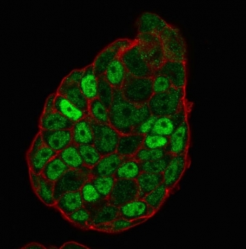
| Applications | Flow cytometry : 1-2ug/10^6 cells Immunofluorescence : 1-2ug/ml Immunohistochemistry (FFPE) : 0.25-0.5ug/ml for 30 min at RT |
| Limitations | This p27Kip1 antibody is available for research use only. |
|
Review this product on BioCompare and get a $20 Amazon gift card
|
Related Products
|
This antibody recognizes a 27kDa protein, identified as the p27Kip1, a cell cycle regulatory mitotic inhibitor. The p27Kip1 antibody is highly specific and shows no cross-reaction with other related mitotic inhibitors. p27Kip1 functions as a negative regulator of G1 progression and has been proposed to function as a possible mediator of TGF beta induced G1 arrest. It is a candidate tumor suppressor gene. This mAb is excellent for staining of formalin-fixed tissues.
The concentration stated for each application is a general starting point. Variations in protocols, secondaries and substrates may require the p27Kip1 antibody to be titered up or down for optimal performance.
1. Staining of formalin-fixed tissues requires boiling tissue sections in 10mM citrate buffer, pH 6.0, for 10-20 min followed by cooling at RT for 20 minutes.
2. The prediluted format is supplied in a dropper bottle and is optimized for use in IHC. After epitope retrieval step (if required), drip mAb solution onto the tissue section and incubate at RT for 30 min.
Purified GST-p27 fusion protein of human origin was used as the immunogen for this p27Kip1 antibody.
Store the p27Kip1 antibody at 2-8oC (with azide) or aliquot and store at -20oC or colder (without azide).

Your bulk quote request has been submitted successfully!
Please contact us if you have any questions.
 Powered by Bioz