- Tel: 858.663.9055
 Email: info@nsjbio.com
- Tel: 858.663.9055
- Email: info@nsjbio.com


| Catalog No | Formulation | Size | Price (USD) | ||
|---|---|---|---|---|---|
 |
V2721-100UG | 0.2 mg/ml in 1X PBS with 0.1 mg/ml BSA (US sourced) and 0.05% sodium azide | 100 ug | 519 | |
|
|
V2721-20UG | 0.2 mg/ml in 1X PBS with 0.1 mg/ml BSA (US sourced) and 0.05% sodium azide | 20 ug | 229 | |
|
|
V2721SAF-100UG | 1 mg/ml in 1X PBS; BSA free, sodium azide free | 100 ug | 519 | |
|
|
V2721IHC-7ML | Prediluted in 1X PBS with 0.1 mg/ml BSA (US sourced) and 0.05% sodium azide; *For IHC use only* | 7 ml | 519 |
| Availability | 1-3 business days |
| Species Reactivity | Human |
| Format | Purified |
| Clonality | Monoclonal (mouse origin) |
| Isotype | Mouse IgG1, kappa |
| Clone Name | HMPV |
| Purity | Protein G affinity chromatography |
| UniProt | P15941 |
| Localization | Cytoplasmic and cell surface |
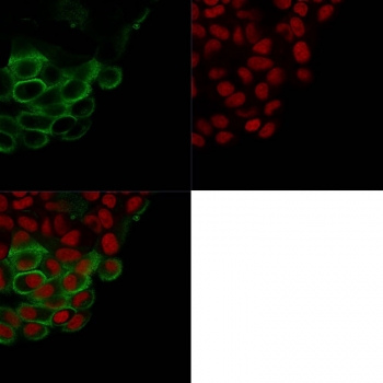
| Applications | Flow cytometry : 1-2ug/10^6 cells Immunofluorescence : 1-2ug/ml Immunohistochemistry (FFPE) : 1-2ug/ml for 30 min at RT Western blot : 1-2ug/ml |
| Limitations | This MUC-1 antibody is available for research use only. |
|
Review this product on BioCompare and get a $20 Amazon gift card
|
Related Products
|
This mAb recognizes full-length MUC1/Mucin-1/Epithelial Marker Antigen/EMA in a glycosylation-independent manner and can bind to the fully glycosylated protein. The dominant epitope of this mAb is APDTR in the VNTR region. It reacts with the core peptide of the MUC1 protein, which is a member of a family of mucin glycoproteins that are characterized by high carbohydrate content, O-linked oligosaccharides, high molecular weight (>200kDa) and an amino acid composition rich in serine, threonine, proline and glycine. The core protein contains a domain of 20 amino-acid tandem repeats that functions as multiple epitopes for the mAb. Incomplete glycosylation of some tumor-associated mucins may lead to variable unmasking of the multiple peptide epitopes leading to the observed differences in staining intensity between normal and malignant tissues. This mAb reacts with both normal and malignant epithelia of various tissues including breast and colon.
Optimal dilution of the MUC-1 antibody should be determined by the researcher.
1. Staining of formalin-fixed tissues requires boiling tissue sections in pH 9 10mM Tris with 1mM EDTA for 10-20 min followed by cooling at RT for 20 min
2. The prediluted format is supplied in a dropper bottle and is optimized for use in IHC. After epitope retrieval step (if required), drip mAb solution onto the tissue section and incubate at RT for 30 min.
Human breast cancer cell line ZR-75 cells were used as the immunogen for the MUC-1 antibody.
Store the MUC-1 antibody at 2-8oC (with azide) or aliquot and store at -20oC or colder (without azide).

Your bulk quote request has been submitted successfully!
Please contact us if you have any questions.
 Powered by Bioz