- Tel: 858.663.9055
 Email: info@nsjbio.com
- Tel: 858.663.9055
- Email: info@nsjbio.com


| Catalog No | Formulation | Size | Price (USD) | ||
|---|---|---|---|---|---|
 |
F53515-0.1ML | In ascites with 0.09% sodium azide | 0.1 ml | 399 |
| Availability | 1-3 business days |
| Species Reactivity | Human |
| Format | Ascites |
| Clonality | Monoclonal (mouse origin) |
| Isotype | Mouse IgG1, k |
| Clone Name | 159CT34.12.3.4 |
| Purity | Ascites |
| UniProt | P11137 |
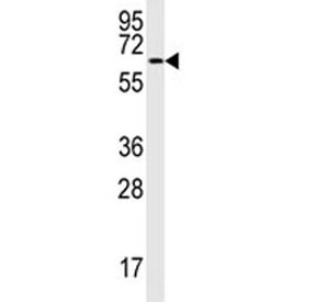
| Applications | Western blot : 1:1000 Immunofluorescence : 1:10-1:50 IHC (Paraffin) : 1:10-1:50 |
| Limitations | This MAP2 antibody is available for research use only. |
|
Review this product on BioCompare and get a $20 Amazon gift card
|
Related Products
|
The exact function of MAP2 is unknown but MAPs may stabilize the microtubules against depolymerization. They also seem to have a stiffening effect on microtubules. [UniProt]
The stated application concentrations are suggested starting amounts. Titration of the MAP2 antibody may be required due to differences in protocols and secondary/substrate sensitivity.
This MAP2 antibody was raised using purified His-tagged recombinant human MAP2.
Aliquot the MAP2 antibody and store frozen at -20oC or colder. Avoid repeated freeze-thaw cycles.

Your bulk quote request has been submitted successfully!
Please contact us if you have any questions.
 Powered by Bioz