- Tel: 858.663.9055
 Email: info@nsjbio.com
- Tel: 858.663.9055
- Email: info@nsjbio.com


| Catalog No | Formulation | Size | Price (USD) | ||
|---|---|---|---|---|---|
 |
R31894 | 0.5mg/ml if reconstituted with 0.2ml sterile DI water | 100 ug | 429 |
| Availability | 1-3 business days |
| Species Reactivity | Human, Mouse, Rat |
| Format | Antigen affinity purified |
| Clonality | Polyclonal (rabbit origin) |
| Isotype | Rabbit IgG |
| Purity | Antigen affinity |
| Buffer | Lyophilized from 1X PBS with 2% Trehalose and 0.025% sodium azide |
| UniProt | P06239 |
| Localization | Cytoplasmic |
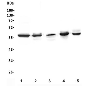
| Applications | Western blot : 0.1-0.5ug/ml Immunohistochemistry (FFPE) : 0.5-1ug/ml Flow cytometry : 1-3ug/10^6 cells Immunocytochemistry : 2-4ug/ml |
| Limitations | This LCK antibody is available for research use only. |
|
Review this product on BioCompare and get a $20 Amazon gift card
|
Related Products
|
Lck (or lymphocyte-specific protein tyrosine kinase) is a protein found inside specialized cells of the immune system called lymphocytes. The human LCK gene is mapped to chromosome 1p35-p32. This gene is a member of the Src family of protein tyrosine kinases (PTKs). The encoded protein is a key signaling molecule in the selection and maturation of developing T-cells. It contains N-terminal sites for myristylation and palmitylation, a PTK domain, and SH2 and SH3 domains which are involved in mediating protein-protein interactions with phosphotyrosine-containing and proline-rich motifs, respectively. The protein localizes to the plasma membrane and pericentrosomal vesicles, and binds to cell surface receptors, including CD4 and CD8, and other signaling molecules. Multiple alternatively spliced variants, encoding the same protein, have been described.
Optimal dilution of the LCK antibody should be determined by the researcher.
Amino acids ELYQLMRLCWKERPEDRPTFDYLRSVLEDFFTATEGQYQ of human Lymphocyte-specific protein tyrosine kinase were used as the immunogen for the LCK antibody.
After reconstitution, the LCK antibody can be stored for up to one month at 4oC. For long-term, aliquot and store at -20oC. Avoid repeated freezing and thawing.

Your bulk quote request has been submitted successfully!
Please contact us if you have any questions.
 Powered by Bioz