- Tel: 858.663.9055
 Email: info@nsjbio.com
- Tel: 858.663.9055
- Email: info@nsjbio.com


| Catalog No | Formulation | Size | Price (USD) | ||
|---|---|---|---|---|---|
 |
V2068-100UG | 0.2 mg/ml in 1X PBS with 0.1 mg/ml BSA (US sourced) and 0.05% sodium azide | 100 ug | 519 | |
|
|
V2068-20UG | 0.2 mg/ml in 1X PBS with 0.1 mg/ml BSA (US sourced) and 0.05% sodium azide | 20 ug | 229 | |
|
|
V2068SAF-100UG | 1 mg/ml in 1X PBS; BSA free, sodium azide free | 100 ug | 519 | |
|
|
V2068IHC-7ML | Prediluted in 1X PBS with 0.1 mg/ml BSA (US sourced) and 0.05% sodium azide; *For IHC use only* | 7 ml | 519 |
| Species Reactivity | Human |
| Format | Purified |
| Clonality | Monoclonal (mouse origin) |
| Isotype | Mouse IgG2a, kappa |
| Clone Name | 156-3C11 |
| Purity | Protein G affinity chromatography |
| Buffer | 1X PBS, pH 7.4 |
| Gene ID | 960 |
| Localization | Cell surface, cytoplasmic |
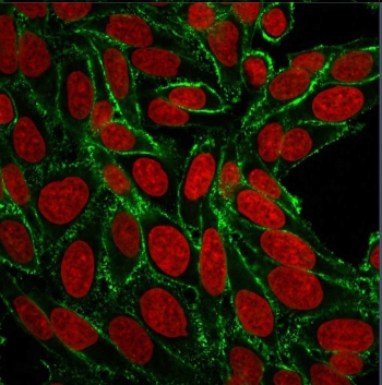
| Applications | Flow cytometry : 2-4ug/10^6 cells Immunofluorescence : 2-4ug/ml Western blot : 1-2ug/ml Immunohistochemistry (FFPE) : 1-2ug/ml for 30 min at RT |
| Limitations | This HCAM antibody is available for research use only. |
|
Review this product on BioCompare and get a $20 Amazon gift card
|
Related Products
|
This antibody recognizes a cell surface glycoprotein of 80-95kDa, called CD44, or HCAM, on lymphocytes, monocytes, and granulocytes (Leucocyte Typing Workshop V). Its epitope is resistant to digestion by trypsin and chymotrypsin. The CD44 family of glycoproteins exists in a number of variant isoforms, the most common being the standard 85-95kDa or hematopoietic variant (CD44-s). Higher molecular weight isoforms are described in epithelial cells (CD44-v), which are believed to function in intercellular adhesion and stromal binding. HCAM antibody immunostaining is commonly used for the discrimination of urothelial transitional cell carcinoma in-situ from non-neoplastic changes in the urothelium.
The concentration stated for each application is a general starting point. Variations in protocols, secondaries and substrates may require the HCAM antibody to be titered up or down for optimal performance.
1. Staining of formalin-fixed tissues requires boiling tissue sections in pH 9 10mM Tris with 1mM EDTA for 10-20 min followed by cooling at RT for 20 minutes.
2. The prediluted format is supplied in a dropper bottle and is optimized for use in IHC. After epitope retrieval step (if required), drip mAb solution onto the tissue section and incubate at RT for 30 min.
Stimulated human leukocytes were used as the immunogen for this CD44 / HCAM antibody.
Store the HCAM antibody at 2-8oC (with azide) or aliquot and store at -20oC or colder (without azide).

Your bulk quote request has been submitted successfully!
Please contact us if you have any questions.
 Powered by Bioz