- Tel: 858.663.9055
 Email: info@nsjbio.com
- Tel: 858.663.9055
- Email: info@nsjbio.com


| Catalog No | Formulation | Size | Price (USD) | ||
|---|---|---|---|---|---|
 |
V2543-100UG | 0.2 mg/ml in 1X PBS with 0.1 mg/ml BSA (US sourced) and 0.05% sodium azide | 100 ug | 519 | |
|
|
V2543-20UG | 0.2 mg/ml in 1X PBS with 0.1 mg/ml BSA (US sourced) and 0.05% sodium azide | 20 ug | 229 | |
|
|
V2543SAF-100UG | 1 mg/ml in 1X PBS; BSA free, sodium azide free | 100 ug | 519 | |
|
|
V2543IHC-7ML | Prediluted in 1X PBS with 0.1 mg/ml BSA (US sourced) and 0.05% sodium azide; *For IHC use only* | 7 ml | 519 |
|
| Availability | 1-3 business days |
| Species Reactivity | Human |
| Format | Purified |
| Clonality | Monoclonal (mouse origin) |
| Isotype | Mouse IgG1, kappa |
| Clone Name | GLG1/970 |
| Purity | Protein G affinity chromatography |
| UniProt | Q92896 |
| Localization | Golgi complex in cytoplasm |
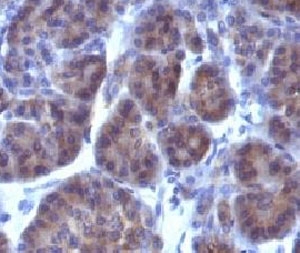
| Applications | Immunohistochemistry (FFPE) : 0.1-0.2ug/ml for 30 min at RT |
| Limitations | This Golgi Marker antibody is available for research use only. |
|
Review this product on BioCompare and get a $20 Amazon gift card
|
Related Products
|
This mAb recognizes a protein of 134kDa, which binds fibroblast growth factor and E-selectin (cell-adhesion lectin on endothelial cells mediating the binding of neutrophils). Fucosylation is essential for binding to E-selectin. It contains sialic acid residues and 16 Cys-rich GLG1 repeats. This mAb can be used to stain the Golgi complex in cell or tissue preparations and can be used as a Golgi marker in subcellular fractions. It produces a diffuse staining pattern of the Golgi zone in normal and malignant cells. This mAb is an excellent marker for human cells in xenographic model research. It reacts specifically with human cells. The Golgi apparatus is an organelle present in all eukaryotic cells that forms a part of the endomembrane system. The primary function of the Golgi apparatus is to process and package macromolecules synthesized by the cell for exocytosis or use within the cell. The Golgi is made up of a stack of flattened, membrane-bound sacs known as cisternae, with three functional regions: the cis face, medial region and trans face. Each region consists of various enzymes that selectively modify the macromolecules passing though them, depending on where they are destined to reside. Several spherical vesicles that have budded off of the Golgi are present surrounding the main cisternae. The Golgi tends to be more pronounced and numerous in cells that make and secrete many substances such as plasma B cells.
Optimal dilution of the Golgi Marker antibody should be determined by the researcher.
1. Staining of formalin-fixed tissues requires boiling tissue sections in pH 9 10mM Tris with 1mM EDTA for 10-20 min followed by cooling at RT for 20 minutes
2. The prediluted format is supplied in a dropper bottle and is optimized for use in IHC. After epitope retrieval step (if required), drip mAb solution onto the tissue section and incubate at RT for 30 min.
The Golgi fraction from human liver cells was used as the immunogen for the Golgi Marker antibody.
Store the Golgi Marker antibody at 2-8oC (with azide) or aliquot and store at -20oC or colder (without azide).

Your bulk quote request has been submitted successfully!
Please contact us if you have any questions.
 Powered by Bioz
