- Tel: 858.663.9055
 Email: info@nsjbio.com
- Tel: 858.663.9055
- Email: info@nsjbio.com


| Catalog No | Formulation | Size | Price (USD) | ||
|---|---|---|---|---|---|
 |
V2282-100UG | 0.2 mg/ml in 1X PBS with 0.1 mg/ml BSA (US sourced) and 0.05% sodium azide | 100 ug | 519 | |
|
|
V2282-20UG | 0.2 mg/ml in 1X PBS with 0.1 mg/ml BSA (US sourced) and 0.05% sodium azide | 20 ug | 229 | |
|
|
V2282SAF-100UG | 1 mg/ml in 1X PBS; BSA free, sodium azide free | 100 ug | 519 | |
|
|
V2282IHC-7ML | Prediluted in 1X PBS with 0.1 mg/ml BSA (US sourced) and 0.05% sodium azide; *For IHC use only* | 7 ml | 519 |
| Species Reactivity | Human |
| Format | Purified |
| Clonality | Monoclonal (mouse origin) |
| Isotype | Mouse IgG2a, kappa |
| Clone Name | BP53-12 |
| Purity | Protein G affinity chromatography |
| Buffer | 1X PBS, pH 7.4 |
| Gene ID | 7157 |
| Localization | Nuclear |
| Applications | Western blot : 1-2ug/ml Immunohistochemistry (FFPE) : 1-2ug/ml for 30 min at RT |
| Limitations | This p53 antibody is available for research use only. |
|
Review this product on BioCompare and get a $20 Amazon gift card
|
Related Products
|
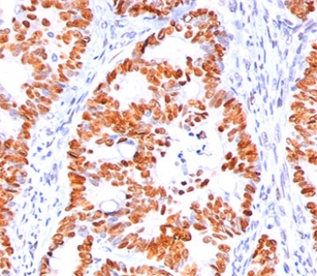
This antibody is specific for a 53kDa protein, which is identified as p53 suppressor gene product. It reacts with the mutant as well as the wild form of p53 under denaturing and non-denaturing conditions. The antibody epitope maps within the N-terminus (aa 20-25) of p53 oncoprotein. p53 is a tumor suppressor gene expressed in a wide variety of tissue types and is involved in regulating cell growth, replication, and apoptosis. It binds to MDM2, SV40 T antigen and human papilloma virus E6 protein. Positive nuclear staining with p53 antibody has been reported to be a negative prognostic factor in breast carcinoma, lung carcinoma, colorectal, and urothelial carcinoma. antibody to p53 positivity has also been used to differentiate uterine serous carcinoma from endometrioid carcinoma as well as to detect intratubular germ cell neoplasia. Mutations involving p53 are found in a wide variety of malignant tumors, including breast, ovarian, bladder, colon, lung, and melanoma.
The concentration stated for each application is a general starting point. Variations in protocols, secondaries and substrates may require the antibody to be titered up or down for optimal performance.
1. Staining of formalin-fixed tissues requires boiling tissue sections in pH 9 10mM Tris with 1mM EDTA for 10-20 min followed by cooling at RT for 20 minutes.
2. The prediluted format is supplied in a dropper bottle and is optimized for use in IHC. After epitope retrieval step (if required), drip mAb solution onto the tissue section and incubate at RT for 30 min.
Recombinant human wild-type p53 protein was used as the immunogen for this antibody.
Store the p53 antibody at 2-8oC (with azide) or aliquot and store at -20oC or colder (without azide).
Antigen NY-CO-13, BCC7, Cellular Tumor Antigen p53, LFS1, TP53, Transformation Related Protein 53 (TRP53), Tumor Protein p53, Tumor Suppressor p53, p53 antibody

Your bulk quote request has been submitted successfully!
Please contact us if you have any questions.
 Powered by Bioz