- Tel: 858.663.9055
 Email: info@nsjbio.com
- Tel: 858.663.9055
- Email: info@nsjbio.com


| Catalog No | Formulation | Size | Price (USD) | ||
|---|---|---|---|---|---|
 |
V2116-100UG | 0.2 mg/ml in 1X PBS with 0.1 mg/ml BSA (US sourced) and 0.05% sodium azide | 100 ug | 519 | |
|
|
V2116-20UG | 0.2 mg/ml in 1X PBS with 0.1 mg/ml BSA (US sourced) and 0.05% sodium azide | 20 ug | 229 | |
|
|
V2116SAF-100UG | 1 mg/ml in 1X PBS; BSA free, sodium azide free | 100 ug | 519 | |
|
|
V2116IHC-7ML | Prediluted in 1X PBS with 0.1 mg/ml BSA (US sourced) and 0.05% sodium azide; *For IHC use only* | 7 ml | 519 |
| Availability | 1-3 business days |
| Species Reactivity | Human |
| Format | Purified |
| Clonality | Monoclonal (mouse origin) |
| Isotype | Mouse IgG2b, lambda |
| Clone Name | M2-7C10 |
| Purity | Protein G affinity chromatography |
| Buffer | 1X PBS, pH 7.4 |
| Gene ID | 2315 |
| Localization | Cytoplasmic |
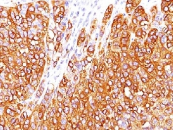
| Applications | Immunohistochemistry (FFPE) : 1-2ug/ml for 30 min at RT Prediluted IHC only format : incubate for 30 min at RT (1) |
| Limitations | This MART-1 antibody is available for research use only. |
|
Review this product on BioCompare and get a $20 Amazon gift card
|
Related Products
|
This antibody recognizes a protein doublet of 20-22kDa, identified as MART-1 (Melanoma Antigen Recognized by T cells 1) or Melan-A. MART-1 is a melanocyte differentiation antigen recognized by autologous cytotoxic T lymphocytes. There are seven other melanoma associated antigens recognized by autologous cytotoxic T cells: MAGE-1, MAGE-3, tyrosinase, gp100, gp75, BAGE-1, and GAGE-1. Subcellular fractionation shows that MART-1 is present in melanosomes and endoplasmic reticulum. This MART-1 antibody labels melanomas and other tumors showing melanocytic differentiation. It is also a useful positive-marker for angiomyolipomas. The antibody does not stain tumor cells of epithelial, lymphoid, glial, or mesenchymal origin.
The concentration stated for each application is a general starting point. Variations in protocols, secondaries and substrates may require the MART-1 antibody to be titered up or down for optimal performance.
1. The prediluted format is supplied in a dropper bottle and is optimized for use in IHC. After epitope retrieval step (if required), drip mAb solution onto the tissue section and incubate at RT for 30 min.
Recombinant human MART-1 protein was used as the immunogen for this antibody.
Store the MART-1 antibody at 2-8oC (with azide) or aliquot and store at -20oC or colder (without azide).
Antigen LB39-AA, Antigen SK29-AA, Melanoma antigen recognized by T-cells 1, MLAN-A, MLANA, MART-1 antibody, Melan-A antibody

Your bulk quote request has been submitted successfully!
Please contact us if you have any questions.
 Powered by Bioz